Illustrating mites
What mites look like under the microscope
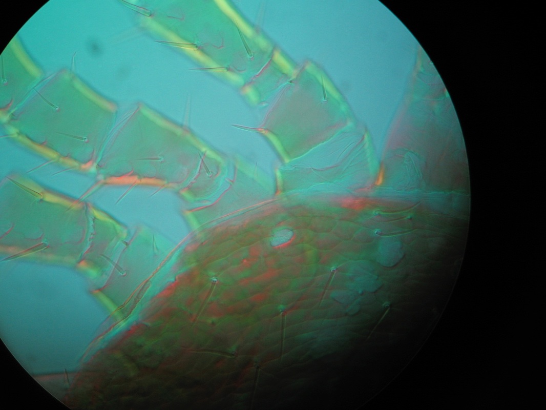
This is the left 'shoulder' and three hairy legs of a mite, as seen at a high magnification. I love the rainbow effect of the special light filter! The mite has been chemically 'cleared', mounted onto a slide and pressed flat under a cover slip.
Drawing with the microscope
|
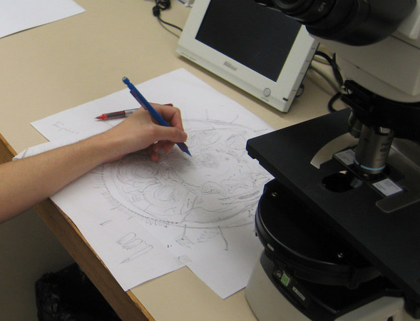
I drew the mites by tracing their features using the camera lucida attachment on the microscope.
Sometimes we'd show features above and below the surface, incorporating as much information as possible into a single picture. This photo shows me drawing the venter (underside) of Berzercon ferdinandi. |
Drawing mites is like drawing a map. The mite is pressed flat onto a slide, and every hair, pore and wrinkle is mapped out in the illustration.
Consulting with the taxonomist
The taxonomist would review my drawing, checking for mistakes like a missing hair or pore. The final drawing is officially filed as the exact description of the species, so it's got to be perfect!
Final illustration in ink
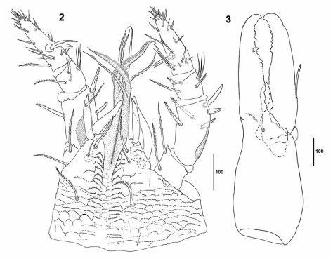
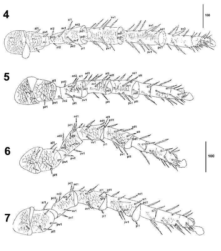
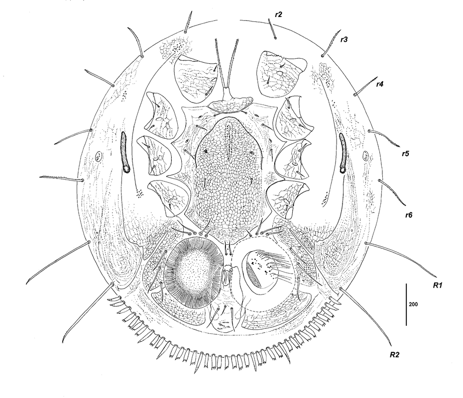
This is the final illustration of the venter of Berzercon ferdinandi.
It's been traced over again in ink, and then digitally labelled for publication.
It's been traced over again in ink, and then digitally labelled for publication.
Berzercon ferdinandi is found on carabid beetles in New Zealand.
Read more about this species on Wikipedia or access the full paper :
Seeman, O.D.; Baker, M.R. 2013: A new genus and species of Discozerconidae (Acari: Mesostigmata) from carabid beetles (Coleoptera: Carabidae) in New Zealand. Zootaxa, 3750(2): 130-142. doi:10.11646/zootaxa.3750.2.2to edit.
Read more about this species on Wikipedia or access the full paper :
Seeman, O.D.; Baker, M.R. 2013: A new genus and species of Discozerconidae (Acari: Mesostigmata) from carabid beetles (Coleoptera: Carabidae) in New Zealand. Zootaxa, 3750(2): 130-142. doi:10.11646/zootaxa.3750.2.2to edit.
Images: All images are illustrated by Michelle Baker, credited to the Queensland Museum.
